Making Of: Preparation Techniques and Photography
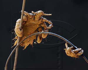
The crab louse as a gymnast? Motif of the 2002 edition.
The images from the LifeSciences calendars 2002 to 2024 come from the scientific photographers Oliver Meckes and Nicole Ottawa, respectively Martin Oeggerli (2014 and 2015 editions). All of our image authors have specialized in the aesthetic representation of scientific facts, with a focus on microscopy.
In 1994 Oliver Meckes and Nicole Ottawa founded the agency "eye of science", Martin Oeggerli has been navigating the microcosm as "Micronaut" since 2008. The most important tools of all three are the scanning electron microscope and powerful computers. At eye of science, whose preparation technique we would like to describe in the following, almost all preparations are produced and photographed in our own SEM laboratory.
Now an example for the creation of such recordings:
The example crab louse
Crab louse in Germany is almost extinct. The Germans are just too clean. Fresh produce from our homeland is therefore not to be expected.
Oliver Meckes and Nicole Ottawa therefore contacted the Tropical Institute in Tübingen. There were actually some specimens there, preserved in 70% alcohol, country of origin Africa. Naturally nice and clean, without hair.
In order to be looked at in the SEM (scanning electron microscope), some preparations were still necessary. The crabs were fixed and transferred step by step into alcohol (ascending row of 70, 80, 90, 95, 99 and 100% of ethanol). That took in the case of the pubic louse 1 day. The preparation was then placed in a pressure chamber, where the alcohol was replaced by liquid carbon dioxide at 50 bar and slowly heated to 40 °C. The liquid carbon dioxide was then removed from the preparation. Finally, the sample was coated with gold in a vacuum.
Only with this complicated process is it possible to ensure that the object does not shrink and deform during drying, or that cells collapse. For the subsequent examination in the vacuum of the SEM, the object must be anhydrous and electrically conductive.
Now there were gold-plated crabs, but they only look lifelike when they are in their millieu. So the animals were put in a wet chamber to make the joints move. Meanwhile Oliver cut some hairs off and sharpened them with the sharpest scissors! The crabs should hang with their legs shaped like snap hooks on the hair like in real life! With a soft special tweezers the crab louse in one hand, with a hard pair of tweezers the hair in the other hand, a half-hour long filament started with 40 times magnification under the stereomicroscope. Thread hair into the crab louse leg. The louse is 2mm long, her leg is just 0.3mm long! After this was successful, the individual hairs had to be "only" placed on a sample holder with the help of electrically conductive adhesive. After another gold sputtering we went to the REM.
And how did the color get into the picture?
Such a SEM, a scanning electron microscope "sees" not with light, but with electrons. Where there is no light, there can be no colours. The electron-optical image can only reflect the topography, not the color of an object defined by light wavelengths. This disadvantage was compensated by eye of science with the help of digital image processing. Nicole Ottawa was patiently busy restoring the color of life to the photographed objects with pixel-precise accuracy (at the time of the photograph everything was gilded). Hair for hair, it is then necessary to separate the object from the background, to give the eyes of a Drosophila their shine back, or to work out the bacteria in the dental plaque or on a small intestine surface cleanly.
Thus, coloring a picture could take 2 days.